

Slicing up the Most Famous Brain
The brain that made the greatest contribution to neuroscience and to our understanding of memory has become a gift that keeps on giving. Patient H.M., or Henry Molaison, gave neuroscience a gift few could imagine: a living map of memory itself. After brain surgery in 1953 to treat epilepsy, he could no longer form new memories—but his cooperation turned his life into a decades-long experiment that revealed the hidden architecture of thought.

Henry Molaison
Surgery and Amnesia
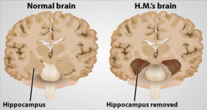
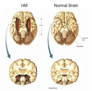
At 27, Molaison underwent removal of parts of his medial temporal lobes, including the hippocampus. His seizures eased, but he could no longer remember new events, and his recall of past experiences became patchy. Researchers observed him for decades, discovering the precise regions of the brain responsible for forming and storing memory.
Memory dissected, literally: Henry Molaison taught us where our past goes when we forget. #neuroscience #memory #neurodope Share on X
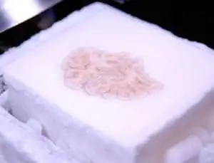
Slicing memory: Each of H.M.’s 2401 brain slices is a window into the architecture of thought.
The Gift of Science
Molaison’s generosity didn’t end with his cooperation. After his death in 2008, his brain was frozen and sliced into 2401 ultra-thin layers. Each slice was photographed, cataloged, and preserved, allowing scientists to reconstruct a digital 3D model of the brain and virtually revisit the surgery that changed neuroscience forever.
Even in death, Patient H.M. keeps schooling scientists on memory. #brainresearch #memorymapping #neurodope Share on X
Slicing memory: Each of H.M.’s 2401 brain slices is a window into the architecture of thought.
A Legacy in Slices
The digital brain model and preserved slices give researchers an unprecedented tool: to trace memory pathways, study epilepsy effects, and test new hypotheses about cognition. Molaison’s brain, meticulously archived, continues to speak, decades later, revealing secrets about how we think, remember, and forget.
Henry Molaison’s brain slices keep whispering the secrets of memory, one virtual cut at a time. #memoryscience #neuroscience #neurodope Share on X
Brain on display: The frozen, dissected brain that revolutionized neuroscience, layer by layer.
Science on Display
The dissection was streamed live, showing the public the painstaking process. Three days, 2401 slices, and infinite lessons. Molaison’s contribution exemplifies the strange intimacy between patient and science, mortality and discovery.
From life to virtual immortality: Patient H.M.’s brain shows that memory is a map we can explore forever. #brainmapping #neurodope #memoryscience Share on X
Legacy of H.M.: Patient and scientist unite in one meticulous experiment that continues to teach us.
When Molaison died of respiratory failure in 2008, he donated his brain to science. That allowed Jacopo Annese at the Brain Observatory in San Diego, California, and his colleagues, to conclusively link his memory problems to specific areas of damaged brain. The dissection was streamed live online.
Molaison is probably the most studied patient in the history of neuroscience, says Annese, and this collection of slices and digital images will provide an unprecedented opportunity for his contribution to neuroscience to continue far beyond his death.

A quick overview of the topics covered in this article.
Latest articles
The Cognitive Rent Economy: How Every App Is Leasing Your Attention Back to You
You don’t lose your attention anymore. You lease it. Modern platforms don’t steal focus. They monetize it, slice it into intervals, and return it [read more...]
AI Is an Opinionated Mirror: What Artificial Intelligence Thinks Consciousness Is
Artificial intelligence thinks it sees us clearly. It does not. It is staring into a funhouse mirror we built out of math, bias, hunger [read more...]

The Age of the Aeronauts: Early Ballon flights
Humans have always wanted to rise above the ground and watch the world shrink beneath them without paying a boarding fee or following traffic [read more...]

Free Energy from the Ether – from Egypt to Tesla
Humans have always chased power from the invisible. From the temples of Egypt to Tesla’s lab in Colorado, inventors sought energy not trapped in [read more...]
The Philosophy of Fake Reality: When Simulation Theory Meets Neuroscience
What if reality isn’t breaking down—but revealing its compression algorithm? Neuroscience doesn’t prove we live in a simulation, but does it show the brain [read more...]
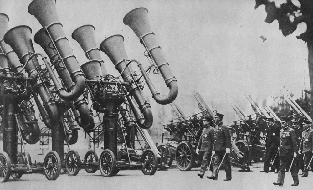
Blast from the Past: Exploring War Tubas – The Sound Locators of Yesteryears
When it comes to innovation in warfare, we often think of advanced technologies like radar, drones, and stealth bombers. However, there was a time [read more...]
Pneumatic Tube Trains – a Lost Antiquitech
Before electrified rails and billion-dollar transit fantasies, cities flirted with a quieter idea: sealed tunnels, air pressure, and human cargo. Pneumatic tube trains weren’t [read more...]

Tartaria and the Soft Reset: The Case for a Quiet Historical Overwrite
Civilizations don’t always collapse with explosions and monuments toppling. Sometimes they dissolve through paperwork, renamed concepts, and smoother stories. Tartaria isn’t a lost empire [read more...]

“A problem cannot be solved by the same consciousness that created it.” – Albert Einstein
“A problem cannot be solved by the same consciousness that created it.” - Albert Einstein
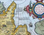
Tartaria: What the Maps Remember
History likes to pretend it has perfect recall, but old maps keep whispering otherwise. Somewhere between the ink stains and the borderlines, a ghost [read more...]


